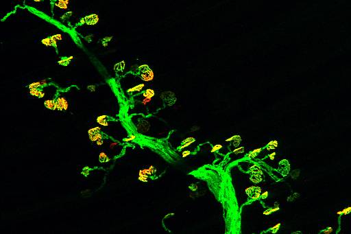
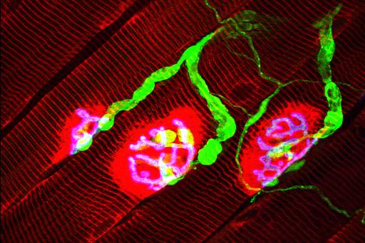
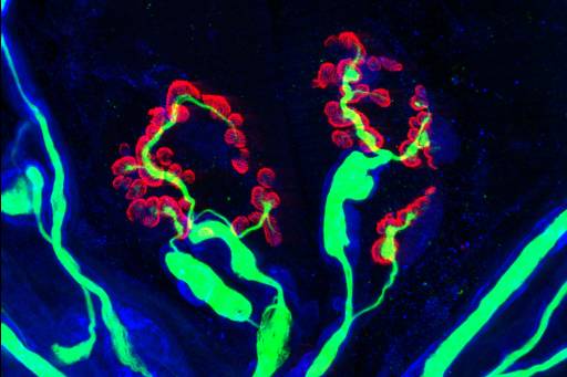
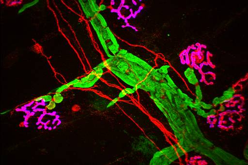
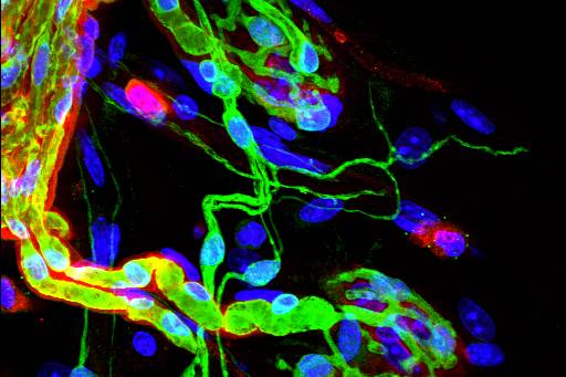
Images from nerve fibers of peripheral nerves; in this region, the glial cell known as Schwann cell associate with the neuron forming around its axon a structure known as the myelin sheath, which increases the propagation velocity of the nerve impulse.
Techniques: Double or triple immunofluorescence staining of teased nerve fibers. Different markers for different compartments of the Schwann cell, axons and basal laminae were used; in some cases, fluorescent-conjugated toxins were used (e.g. rhodamine-phalloidin). Images were taken by laser confocal microscopy and spinning disk laser microcopy. Full maximun projections are shown. Images taken by Felipe Court.